
The human eye, this marvel of biological evolution, operates like a living camera within our body. By examining its complex structure, we discover a perfect harmony among its various components. The eyelids, protective guardians, open onto the eyeball, where the curved cornea collaborates with the lens to focus light. This light then travels through the vitreous before reaching the retina, this sensitive screen where the image is formed. Here, photoreceptor cells and neurons intertwine to transmit visual information to the brain, a process that allows us to perceive the world in all its splendor.
Structure and Functioning of the Eye: From Light to Perception
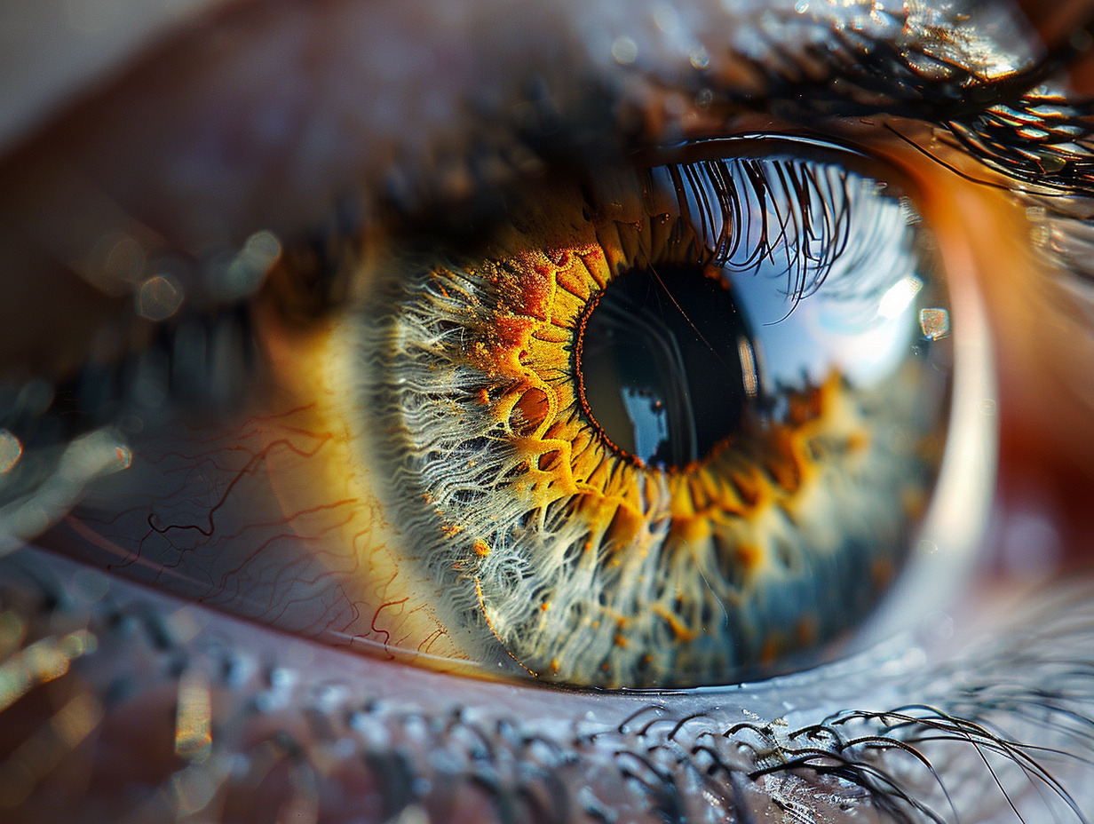
The ocular anatomy reveals itself in all its complexity when considering the eyeball, a spherical organ with an average length of 24 mm, weighing about 7 g and having a volume of 6.5 cm³. This organ, located in the orbit, is responsible for visual function, a feat that begins with the capture of photons. Light first enters through the cornea, this transparent anterior part of the eyeball, before passing through the aqueous humor, a transparent medium contained within the anterior segment. Then, it encounters the iris, this pigmented and contractile membrane, which adjusts the size of the pupil and thus regulates the amount of light reaching the lens. The lens, an essential refractive element, concentrates light rays onto the retina, thanks to its accommodation ability, changing its curvature for precise focusing. Within the retina, photoreceptors transform light signals into electrical impulses, which are then transmitted to the optic nerve. The ciliary body, part of the uvea located behind the iris, plays a fundamental role in this accommodation by adjusting the shape of the lens via its ciliary muscles. Beyond the lens, the vitreous humor, a gel-like substance, fills the posterior segment and maintains the spherical shape of the eye while allowing light to reach the retina. The protection of this refined optical system is ensured by the sclera, the outer covering of the eyeball, as well as by the conjunctiva, a transparent mucous membrane that covers it. The conjunctiva also forms the ‘conjunctival sac’, a pocket between the eyelid and the sclera, which allows for free movement of the eye and the even distribution of the tear film. The choroid, located between the retina and the sclera, provides the necessary nutrition to the eye. The iridocorneal angle ensures the drainage of the aqueous humor, thus preserving intraocular pressure and the health of the eye.
See also : The subtleties of a vehicle's fiscal power: from theory to practice
Protection and Maintenance of the Eye: Eyelids, Tears, and Care
The eyelids play a crucial role in protecting the visual apparatus. They cover the anterior part of the eyeball, moving thanks to the extraocular muscles that orchestrate their opening and closing. This incessant ballet contributes to the distribution of the tear film, essential for the lubrication and hydration of the ocular surface. The eyelids also act as physical barriers against impurities and potential injuries. The lacrimal system, for its part, ensures the production and drainage of tears. These tears, beyond their emotional role, form a protective film on the cornea, providing nutrients and oxygen while eliminating debris. Tear secretion constantly adapts to the environment, increasing in response to irritation or the need for additional hydration. The lacrimal ducts, at the end of their journey, direct tears to the nose, where they are eliminated or reabsorbed. A rigorous visual hygiene is essential for preserving ocular function. Daily care, including proper eyelid cleaning and protection against ultraviolet radiation, helps prevent infections and diseases. Health professionals also recommend moderating exposure to bright screens, taking regular breaks during prolonged visual activities, and adopting adequate lighting to maintain eye health. Paying attention to symptoms such as dry eyes, redness, or visual fatigue invites consultation with an ophthalmologist. Timely diagnosis of conditions such as Claude Bernard-Horner syndrome, characterized by eyelid asymmetry and abnormal pupillary reaction, allows for early treatment and avoids complications. Monitoring and eye care are therefore essential preventive measures to maintain optimal vision.
See also : Practical guide to accessing the Arena portal of the Amiens academy from home